missing translation for 'onlineSavingsMsg'
Learn More
Learn More
Invitrogen™ CD4 Monoclonal Antibody (SK3 (SK-3)), NovaFluor™ Violet 745, eBioscience™
Mouse Monoclonal Antibody
Brand: Invitrogen™ H001T03V02-A
This item is not returnable.
View return policy
Description
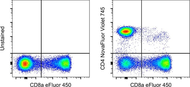
Description: The SK3 monoclonal antibody reacts with human CD4, a 59-kDa cell surface receptor expressed by a majority of thymocytes, a subpopulation of mature T helper cells, and at low levels on monocytes. CD4 is a receptor for the human immunodeficiency virus (HIV). SK3 blocks HIV binding and mixed lymphocyte reaction. The SK3 and RPA-T4 monoclonal antibodies do not cross-block binding, suggesting recognition of distinct epitopes. This product contains 1 vial of NovaFluor conjugate and 1 vial of CellBlox Plus Blocking Buffer. Applications Tested: This SK3 (SK-3) antibody has been pre-titrated and tested by flow cytometric analysis of normal human peripheral blood cells. This can be used at 5 μL (0.1 μg) per test. A test is defined as the amount (μg) of antibody that will stain a cell sample in a final volume of 100 μL. Cell number should be determined empirically but can range from 10^5 to 10^8 cells/test. Master mixes: Master mixes of NFs should be made at 2-8 °C and may be made up to 4 hours ahead of time. We do not recommend storing master mixes containing NovaFluor conjugates overnight or longer. Whole Blood compatibility: When utilizing whole blood (as opposed to density-gradient-purified PBMC), we recommend lysing red blood cells in bulk prior to staining with NovaFluor conjugates.
The CD4 antigen is involved in the recognition of MHC class II molecules and is a co-receptor for HIV. CD4 is primarily expressed in a subset of T-lymphocytes, also referred to as T helper cells, but may also be expressed by other cells in the immune system, such as monocytes, macrophages, and dendritic cells. At the tissue level, CD4 expression may be detected in thymus, lymph nodes, tonsils, and spleen, and also in specific regions of the brain, gut, and other non-lymphoid tissues. CD4 functions to initiate or augment the early phase of T-cell activation through its association with the T-cell receptor complex and protein tyrosine kinase, Lck. It may also function as an important mediator of direct neuronal damage in infectious and immune-mediated diseases of the central nervous system. Multiple alternatively spliced transcripts have been identified in this gene [RefSeq, July 2017].Specifications
| CD4 | |
| Monoclonal | |
| 0.1 μg/Test | |
| PBS with BSA and 0.09% sodium azide; pH 7.2 | |
| P01730 | |
| CD4 | |
| Affinity chromatography | |
| RUO | |
| 920 | |
| 4°C, store in dark, DO NOT FREEZE! | |
| Liquid |
| Flow Cytometry | |
| SK3 (SK-3) | |
| NovaFluor Violet 745 | |
| CD4 | |
| Activation B7-1 antigen; B7; B7.1; B7-1; BB1; B-lymphocyte activation antigen B7; CD28LG; CD28LG1; CD4; CD4 antigen; CD4 antigen (p55); CD4 antigen p55; Cd4 molecule; CD4 precursor; CD4 receptor; CD4, allele 1; cd4a; CD4mut; CD80; CD80 antigen (CD28 antigen ligand 1, B7-1 antigen); CD80 molecule; cell surface glycoprotein CD4; costimulatory factor CD80; costimulatory molecule variant IgV-CD80; CTLA-4 counter-receptor B7.1; fCD4; L3T4; LAB7; Leu-3; Ly-4; lymphocyte antigen CD4; lymphocyte antigen CD4 precursor; membrane protein; p55; T-cell differentiation antigen L3T4; T-cell surface antigen T4/Leu-3; T-cell surface glycoprotein CD4; T-cell surface glycoprotein CD4 precursor (T-cell surface antigen T4/Leu-3) (T-cell differentiation antigen L3T4); T-lymphocyte activation antigen CD80; W3/25; W3/25 antigen | |
| Mouse | |
| 100 Tests | |
| Primary | |
| Human | |
| Antibody | |
| IgG1 κ |
Product Content Correction
Your input is important to us. Please complete this form to provide feedback related to the content on this product.
Product Title
Spot an opportunity for improvement?Share a Content Correction